Ultrasonographic findings in dogs diagnosed with hyperadrenocorticism – retrospective study (2013 to 2020)
DOI:
https://doi.org/10.21708/avb.2022.16.1.10405Resumo
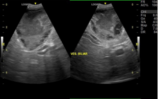
Hyperadrenocorticism is a relatively common endocrinopathy in middle-aged and older dogs, which has become increasingly frequent in the clinical routine. The diagnosis is made by information obtained by history, physical exam findings and results of screening and specific endocrine tests. In addition to laboratory tests, imaging diagnosis, such as ultrasound, can aid on evaluation of possible changes in these cases; also, they may reveal the possible involvement of other organs and systems. The aim of this study was to present the main ultrasonographic changes observed in 18 dogs with hyperadrenocorticism, diagnosed from 2013 to 2020 by the low-dose dexamethasone suppression test, among which hepatomegaly, splenomegaly, gallbladder sludge, renal changes, cystitis and changes in adrenal size, such as bilateral and/or unilateral adrenomegaly, can be cited. Ultrasound examination, therefore, is an examination that can help the clinician in confirming the diagnosis of hyperadrenocorticism, in addition to allowing differentiation between pituitary-dependent hyperadrenocorticism (PDH) and adrenal tumors, and the evaluation of possible secondary changes to the disease.
Downloads
Downloads
Publicado
Edição
Seção
Licença
Autores que publicam na Acta Veterinaria Brasilica concordam com os seguintes termos: a) Autores mantém os direitos autorais e concedem à revista o direito de primeira publicação, com o trabalho simultaneamente licenciado sob a Licença Creative Commons Attribution que permite o compartilhamento do trabalho com reconhecimento da autoria e publicação inicial nesta revista. b) Autores têm autorização para assumir contratos adicionais separadamente, para distribuição não-exclusiva da versão do trabalho publicada nesta revista (ex.: publicar em repositório institucional ou como capítulo de livro), com reconhecimento de autoria e publicação inicial nesta revista. c) Autores têm permissão e são estimulados a publicar e distribuir seu trabalho online (ex.: em repositórios institucionais ou na sua página pessoal) a qualquer ponto antes ou durante o processo editorial, já que isso pode gerar alterações produtivas, bem como aumentar o impacto e a citação do trabalho publicado (Veja O Efeito do Acesso Livre).

 Esta obra está licenciada com uma Licença